
Aggression is often associated as a negative emotion. Uncontrolled aggression can lead to conflict, violence and negative consequences for individuals and society. Yet that does not that mean that aggression serves no purpose. It is an instinctive behavior found in many species that may be necessary for survival. The key is managing and channeling aggression.
In a recent study using mice, researchers at Tohoku University have demonstrated that neuron-glial interactions in the cerebellum set the tone of aggression, suggesting that future therapeutic methods could rely on adjusting glial activity there to manage anger and aggression.
The findings were detailed in the journal, Neuroscience Research, on November 24, 2023.
Scientists have recently recognized the role of the cerebellum in non-motor functions such as social cognition. A malfuctioning cerebellum can occur in autism spectrum disorders and schizophrenia, leading to social interaction difficulties. In particular, it has been reported that a region of the cerebellum, known as the vermis, is associated with aggression in humans. Therefore, the researchers investigated the possibility that Bergmann glial cells in the cerebellar vermis regulate the volume of aggression in mice.
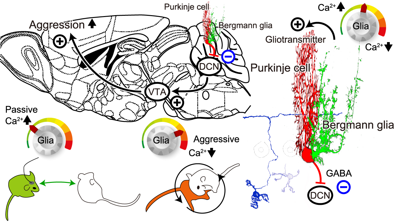
Schematic diagram of the mechanism of cerebellar glial cell activity in regulating aggression.
Gliotransmitter could be released in response to intracellular Ca2+ concentration increase in the cerebellar Bergmann glial cells. Excitatory transmitters such as glutamate would increase the cerebellar Purkinje cell activity. The only output nerve fiber from the cerebellum is the axon of the Purkinje cell (PC). PC activity leads to inhibitory GABA input to the deep cerebellar nuclei (DCN). Excitatory neuronal contacts from the DCN to the ventral tegmental area (VTA) have recently been identified. Dopaminergic neurons in the VTA have been reported to have a significant influence on social behavior, including aggression. Therefore, if the schematic pathway as described works, then an increase in Ca2+ concentration in cerebellar glial cells would be expected to increase PC activity and suppress DCN and VTA activity, leading to reduced aggression. This could lead to an early breakup of fights. On the other hand, a decrease in Ca2+ concentration in cerebellar glial cells would decrease PC activity and increase DCN and VTA activity, which would increase aggression and lead to dominance in fight situations. ©Yuki Asano, Ko Matsui
“Cells in the brain can be divided into neurons and glia, and although glia occupy approximately half of the brain, their participation in the brain’s information processing, plasticity, and health has long been an enigma,” says Professor Ko Matsui of the Super-network Brain Physiology lab at Tohoku University, who led the research. “Our newly created fiber photometry method provides a gateway for understanding the physiology of glia.”
Matsui and his colleagues employed the resident-intruder model, where one mouse (the intruder) goes into the territory of another mouse (the resident). When the unfamiliar male mouse enters the cage, quite commonly, a series of fights break out between the resident male mouse and the intruder. Each combat round lasted about 10 seconds, and these rounds were repeated at a frequency of approximately one per minute. The superiority and inferiority of the resident and intruder dynamically switched within each combat round.
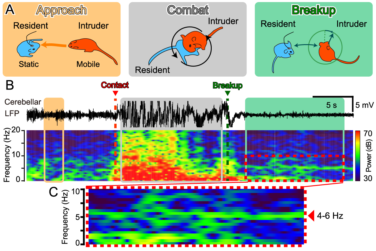
Theta band local field potential (LFP) in the cerebellum upon combat breakup. LFPs were recorded between two electrodes implanted in the mouse cerebellum. In the particular combat round shown, the intruder mouse approached the resident mouse (the recorded mouse), and a fight broke out immediately after contact. Signals recorded during the combat were not analyzed as they contained electrical signals from muscle movements from intense exercise. After the combat breaks up, both mice became stationary. The LFPs in the cerebellum showed oscillations in the frequency range of 4 - 6 Hz (theta band). In separate experiments, when theta band electrical stimulation was delivered via the same cerebellar electrode immediately after the start of the fight, the combat often broke up quickly. It was also shown that ChR2 photostimulation of cerebellar glial cells results in theta band LFP in the cerebellum and also causes the fight to break up early. ©Yuki Asano, Ko Matsui
The fiber photometry method revealed that intracellular Ca2+ levels in cerebellar glia decreased or increased in conjunction with the superiority or inferiority of the fight, respectively. When the combat broke up, the researchers observed 4 to 6 Hz theta band local field potentials in the cerebellum, along with a sustained increase in Ca2+ levels in the glia. Optogenetic stimulation of cerebellar glia induced the emergence of the theta band, casuing an early breakup of the fighting.
Glia have been shown to control the local ionic and metabotropic environment in the brain and also to release transmitters that can affect neuronal activity in the vicinity. The results of this study suggest that the theta band cerebellar neuronal activity is regulated by the activity of Bergmann glial cells, thereby demonstrating that cerebellar glial cells play a role in regulating aggression in mice.
Lead study investigator, Yuki Asano, says that future anger management strategies and clinical control of excessive aggression and violent behavior may be realized by developing a therapeutic strategy that adjusts glial activity in the cerebellum. “Imagine a world without social conflict. By harnessing the innate ability of the cerebellar glia to control aggression, the peaceful future could be become reality.”
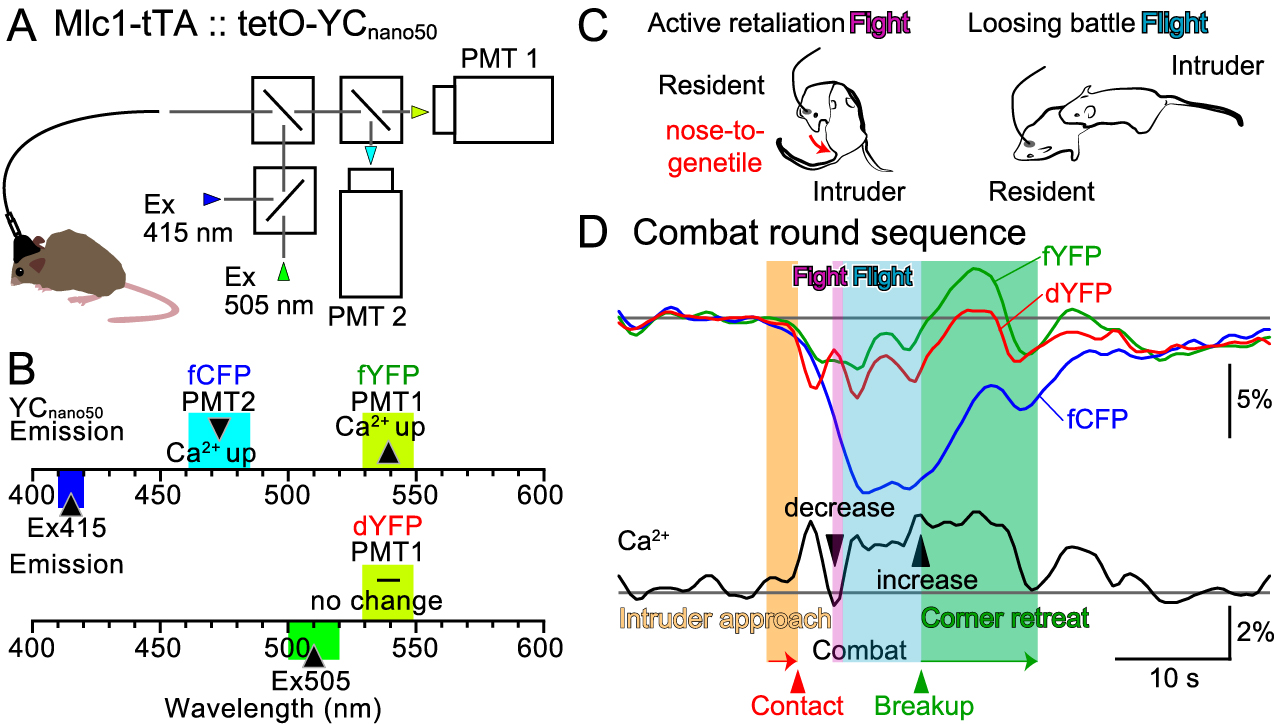
Fiber optic measurement of the local brain environment of the cerebellum. We measured three types of fluorescence (fYFP, dYFP, and fCFP) by inserting an optical fiber into the cerebellum and sending two types of excitation light. By comparing the three fluorescence waveforms, we can estimate the fluctuation of Ca2+ concentration in glial cells, pH fluctuation, and local brain blood volume. Analysis of Ca2+ concentration fluctuations in glial cells during combats revealed that Ca2+ levels decreased significantly when the recorded resident mice fought back and attacked the intruder mice. When the recorded mice were chased from behind by the intruder mice, Ca2+ in the glial cells increased significantly. In addition, the Ca2+ levels remained high even after the fight broke up. These results suggest that cerebellar glial cell activity is linked to mouse aggression. ©Yuki Asano, Ko Matsui
Publication Details:
Title: Glial tone of aggression
Authors: Yuki Asano, Daichi Sasaki, Yoko Ikoma, Ko Matsui
Journal: Neuroscience Research
DOI: https://doi.org/10.1016/j.neures.2023.11.008
Authors: Yuki Asano, Daichi Sasaki, Yoko Ikoma, Ko Matsui
Journal: Neuroscience Research
DOI: https://doi.org/10.1016/j.neures.2023.11.008
Contact:
Ko Matsui,
Super-network Brain Physiology, Graduate School of Life Sciences,
Tohoku University
Email: matsui@med.tohoku.ac.jp
Website: http://www.ims.med.tohoku.ac.jp/matsui/
Ko Matsui,
Super-network Brain Physiology, Graduate School of Life Sciences,
Tohoku University
Email: matsui@med.tohoku.ac.jp
Website: http://www.ims.med.tohoku.ac.jp/matsui/