
Researchers at Tohoku University have shown that astrocytes - star-shaped glial cells that control the local ionic and metabotropic environment of the brain - exhibit an acid response with REM sleep in mice. They theorize that the acid response could be the underlying drive for specific information processing and generating plasticity during sleep.
They further discovered that REM response in astrocytes intensified in the epileptic brain, meaning studying brain environmental changes associated with REM sleep could potentially be employed as a biomarker for the severity of epileptogenesis.
The findings were detailed in the journal Brain on March 3, 2023.
Neurons are undoubtedly responsible for information processing in the brain. Astrocytes were not thought to be an essential component of the neural information circuit. However, recent findings suggest that the state of the mind, such as consciousness, sleep, memory formation, and meta-plasticity may all be controlled by astrocytes’ actions.
To understand the role of astrocytes in brain function, fluorescent sensor proteins were genetically expressed in the astrocytes of mice. The researchers implanted an optical fiber into the mice’s lateral hypothalamus, a part of the brain vital for controlling our state of being asleep or awake and whole-body metabolism.
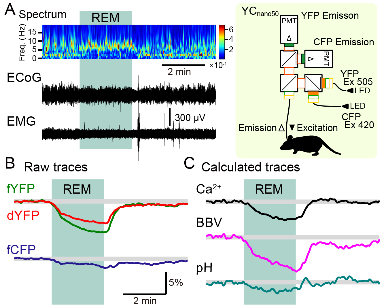
Estimating brain environmental changes from optical signals detected during REM sleep.
(A) Potentials recorded at the cerebral cortex (ECoG) and electromyograms (EMG) were recorded from experimental mice to identify the timing of REM sleep (left). At the same time, an optical fiber was inserted into the lateral hypothalamus and three different fluorescence waveforms (fCFP, fYFP, dYFP) were measured (right; fiber photometry method).
(B) Fluorescence waveforms recorded from the lateral hypothalamus of mice genetically expressing FRET-type calcium (Ca2+) sensor protein in astrocytes. When Ca2+ increases, fCFP (blue fluorescence excited with purple) is expected to decrease and fYFP (yellow fluorescence excited with purple) is expected to increase. dYFP (direct yellow fluorescence excited with green) is considered to remain constant regardless of the Ca2+ concentration. In the present study, fYFP and fCFP did not show a mirror image reaction with REM sleep. In addition, dYFP fluorescence also changed, suggesting that factors other than Ca2+ that affect fluorescence has also changed.
(C) When the intracellular pH becomes acidic, the fluorescence of both fYFP and dYFP decreases, while fCFP is expected to be less affected. In addition, if the vascular diameter near the optical fiber tip swells, an increase in the local blood flow occurs resulting in the fluorescent protein-free area to expand. When such an event occurs, the fluorescence of fYFP, dYFP, and fCFP are all expected to decrease. Considering these effects, we developed a method to extract changes in astrocyte intracellular Ca2+, pH, and local blood flow from the three measured fluorescence waveforms (fYFP, dYFP, and fCPF). During REM sleep, intracellular Ca2+ in astrocytes was shown to decrease, local blood flow (BBV) increased (indicated in the figure to reflect increased blood flow when the waveform shows downward deflection), and intracellular pH in astrocytes became more acidic. The astrocyte acidification in particular was unexpected. This is the first example showing that intracellular pH can fluctuate even under physiological conditions, despite the fact that intracellular pH is strongly buffered with bicarbonate.©Yoko Ikoma & Ko Matsui
Excitation light was sent through this fiber and the emitted fluorescence signals were recorded. Using a newly devised method, the researchers dissected the calcium concentration and pH of the astrocytes and the local brain blood volume changes from the recorded optical signals.
A clear change in the optical signals associated with REM sleep was observed. A calcium decrease, pH decrease (i.e. acidification), and increase in local brain blood volume occurred. The researchers identified that acidification and blood volume changes produce a strong effect on the optical signals; thus, many of the previous studies using fiber photometry could have misinterpreted their recorded data.
Acidification was especially unexpected, as the intracellular solution of cells is highly buffered for pH. Strong acidification occurs upon ischemia but changes in pH were not assumed to occur under physiological conditions. This astrocyte acidification may drive the amplification of synaptic signals and may underlie memory formation during REM sleep.
Interestingly, changes in the local brain environment detected with the optical recordings preceded the signature change of the ensemble neuronal electrical activity detected with electroencephalogram by nearly 20 seconds. This suggests that astrocytes and vascular changes have control of the state of neuronal activity. Transition to REM sleep can also be predicted from these local brain environmental changes.
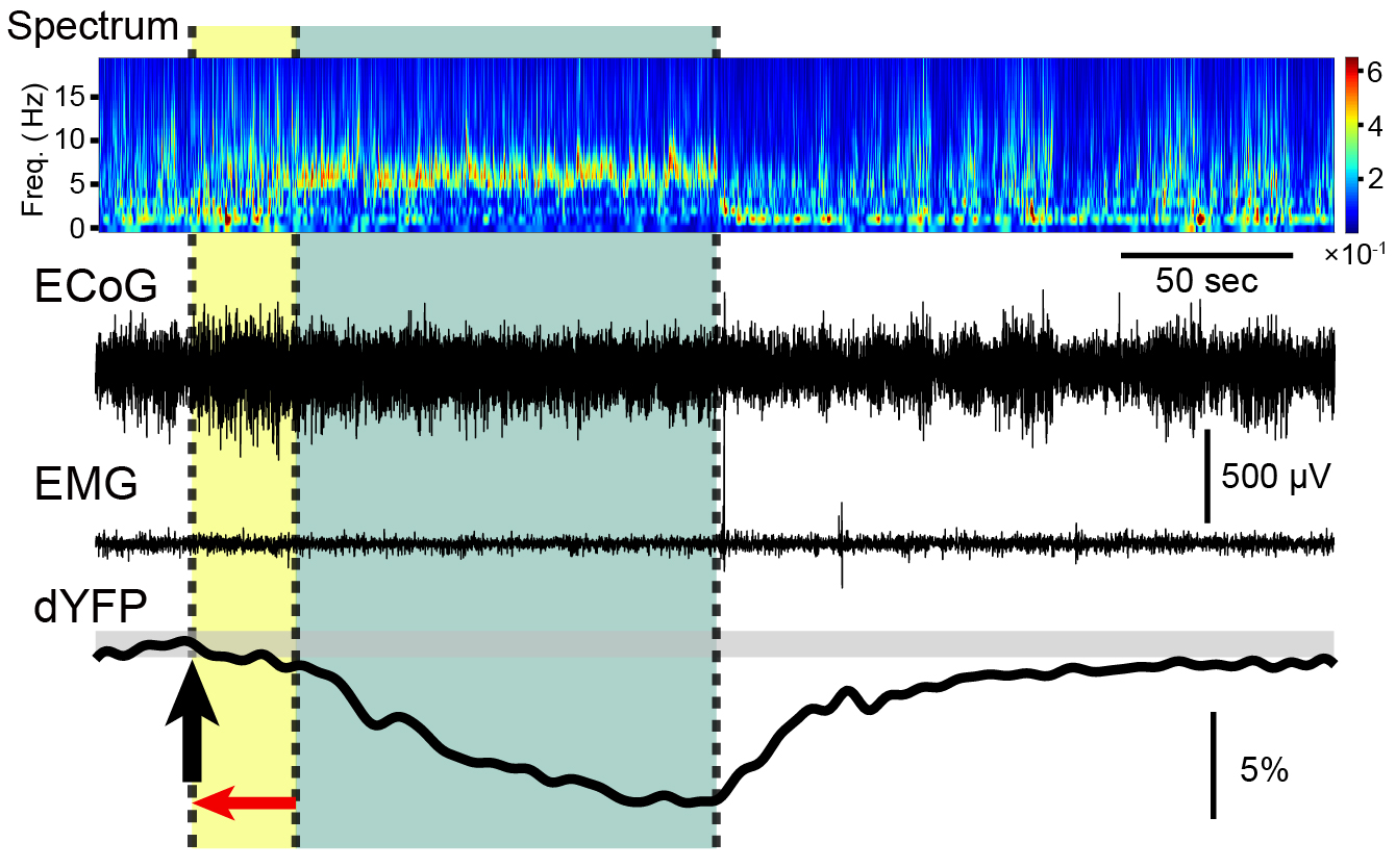
Brain environment change precedes REM sleep transition.
By analyzing the frequency of the electroencephalogram (ECoG), it is possible to determine when the theta waves (6 - 9 Hz) increase and the delta waves (1 - 4 Hz) decrease, indicating the transition to REM sleep. On the other hand, a close examination of the timing of the downward deflection in dYFP fluorescence recorded by fiber photometry in the lateral hypothalamus reveals that it precedes the onset of REM sleep determined by ECoG by nearly 20 seconds. The measured fluorescence emitted from the dYFP is thought to decrease as the pH in astrocytes becomes more acidic and local blood volume (BBV) increases. These changes in the brain environment precede changes in brain neural activity, indicating that changes in astrocyte and vascular activity cooperate with neurons to influence brain functions such as REM sleep.©Yoko Ikoma & Ko Matsui
By analyzing the frequency of the electroencephalogram (ECoG), it is possible to determine when the theta waves (6 - 9 Hz) increase and the delta waves (1 - 4 Hz) decrease, indicating the transition to REM sleep. On the other hand, a close examination of the timing of the downward deflection in dYFP fluorescence recorded by fiber photometry in the lateral hypothalamus reveals that it precedes the onset of REM sleep determined by ECoG by nearly 20 seconds. The measured fluorescence emitted from the dYFP is thought to decrease as the pH in astrocytes becomes more acidic and local blood volume (BBV) increases. These changes in the brain environment precede changes in brain neural activity, indicating that changes in astrocyte and vascular activity cooperate with neurons to influence brain functions such as REM sleep.©Yoko Ikoma & Ko Matsui
“During REM sleep, prior experiences are sorted and remembered or forgotten, and this process is likely perceived as dreams,” says Professor Ko Matsui of the Super-network Brain Physiology lab at Tohoku University, who led the research. “Acidification in astrocytes may control the likelihood of plasticity to occur in the neural circuits.”
The researchers further studied how the properties of REM sleep change with epilepsy. Repeated stimulus to a mouse’s hippocampus produces a brain prone to hyperactivity and this “kindling” method has been used as a model of epileptogenesis. After kindling, spontaneously occurring REM sleep episodes were recorded. Surprisingly, very little astrocytic calcium decreases and local brain blood volume increases occurred with REM sleep, and a strong acid response was recorded from astrocytes.
“Our previous study has shown an increased acid response of astrocytes associated with intensified epileptic seizures,” says the lead study investigator, Dr. Yoko Ikoma. “Information is transmitted and processed with electrical signals in neurons. The pH of astrocytes may have control over these neuronal activities both in physiology and in disease.”
Monitoring of the bulk pH and local brain blood flow is possible in humans using fMRI. Ikoma says that these local brain environmental changes associated with REM sleep can potentially be used to diagnose the severity of epilepsy in human patients. “A therapeutic strategy designed to control astrocytes’ pH could potentially be used for preventing exacerbation of epilepsy.”
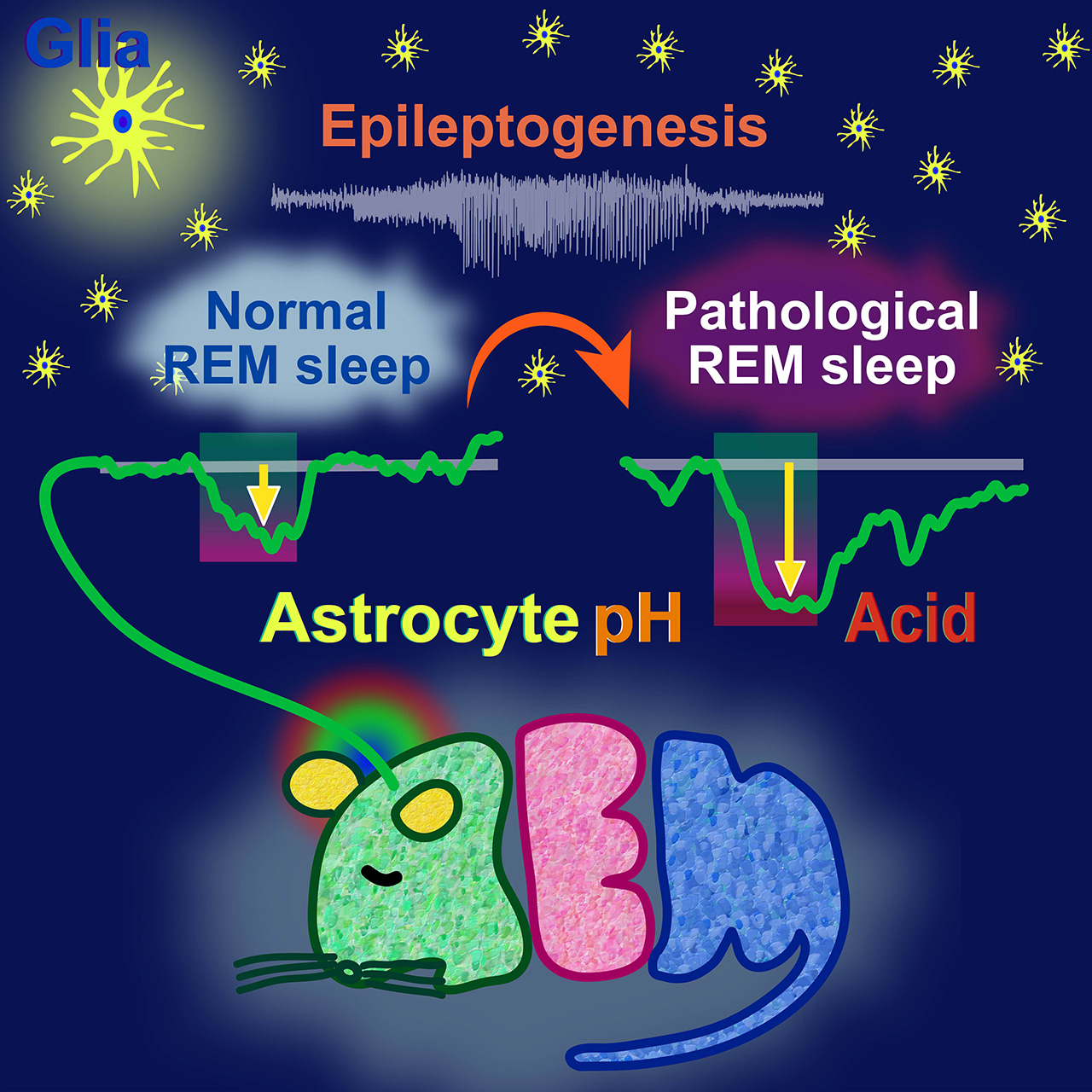
Caption: Acidification of astrocytes in the lateral hypothalamus during REM sleep is enhanced in the epileptic pathological brain.
Analysis of fluorescent waveforms obtained by inserting an optical fiber into the lateral hypothalamus of mice revealed that acidification in astrocytes occurs during REM sleep. Repeated daily electrical stimulation of the hippocampus changes the brain into one prone to epileptic seizures. We analyzed the spontaneous REM sleep that occurs after the brain is transformed into an epileptic pathological brain. The results revealed that astrocytes become more acidic during REM sleep. By analyzing the environmental changes in the brain during REM sleep, it is hoped that a new strategy to diagnose the developmental level of epilepsy and to treat epilepsy pathology by controlling astrocyte acidity can be pioneered.©Yoko Ikoma & Ko Matsui
Analysis of fluorescent waveforms obtained by inserting an optical fiber into the lateral hypothalamus of mice revealed that acidification in astrocytes occurs during REM sleep. Repeated daily electrical stimulation of the hippocampus changes the brain into one prone to epileptic seizures. We analyzed the spontaneous REM sleep that occurs after the brain is transformed into an epileptic pathological brain. The results revealed that astrocytes become more acidic during REM sleep. By analyzing the environmental changes in the brain during REM sleep, it is hoped that a new strategy to diagnose the developmental level of epilepsy and to treat epilepsy pathology by controlling astrocyte acidity can be pioneered.©Yoko Ikoma & Ko Matsui
Publication Details:
Title: Properties of REM sleep alterations with epilepsy
Authors: Yoko Ikoma, Yusuke Takahashi, Daichi Sasaki, Ko Matsui
Journal: Brain
DOI: doi.org/10.1093/brain/awac499
Authors: Yoko Ikoma, Yusuke Takahashi, Daichi Sasaki, Ko Matsui
Journal: Brain
DOI: doi.org/10.1093/brain/awac499
Contact:
Name: Ko Matsui
Affiliation: Super-network Brain Physiology, Graduate School of Life Sciences, Tohoku University
Email: matsui@med.tohoku.ac.jp
Website: http://www.ims.med.tohoku.ac.jp/matsui/
Affiliation: Super-network Brain Physiology, Graduate School of Life Sciences, Tohoku University
Email: matsui@med.tohoku.ac.jp
Website: http://www.ims.med.tohoku.ac.jp/matsui/